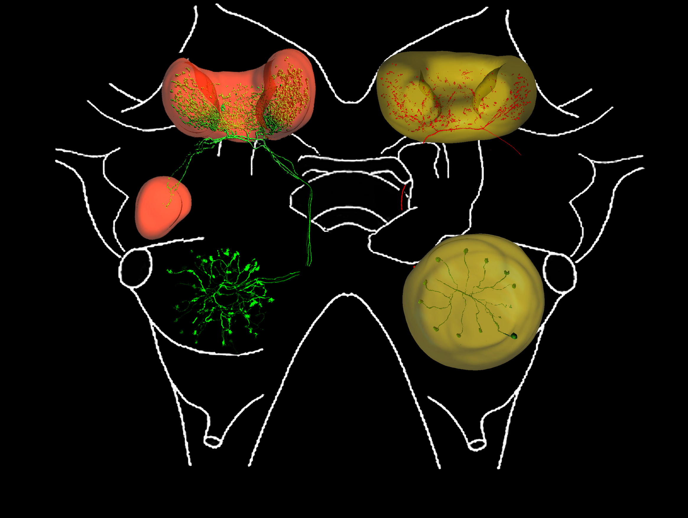
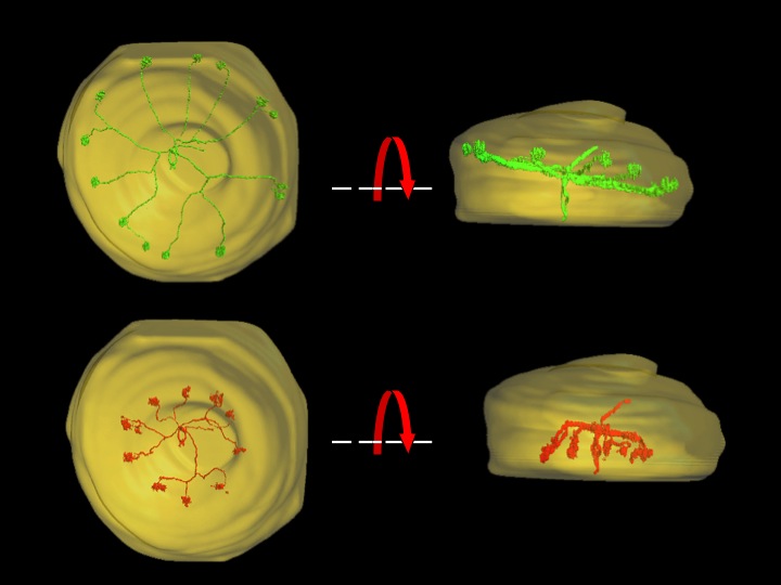
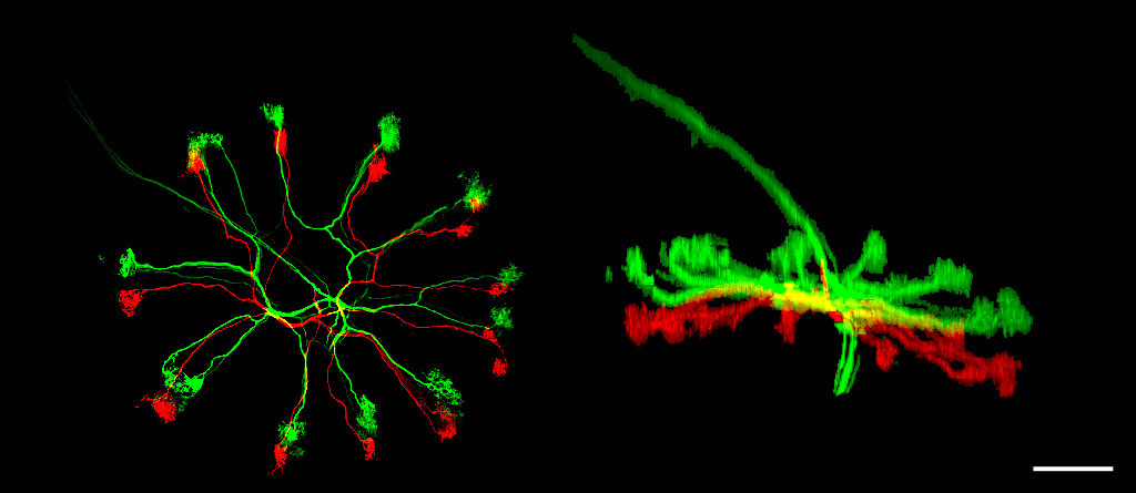
Back

Projection Neurons
Copyright S Sarah Farivar-Hayes, 2022.


PNs IN THE WHOLE BRAIN
Left side of locust brain shows a group of 3 PNs (green) stained together, with their sonata and glomerular projections within the AL, and their projections up to the calyx of the MB and the Lateral Horn (LH). MB calyx and LH are rendered in red. The right side of the brain shows a single PN (green) within the AL (rendered as a yellow sphere), and its projections up to the MB calyx (also rendered in yellow).


PNs IN THE AL
Two different PNs (red and green) shown within the AL (yellow). The images on the left side show the ventral aspect, while those on the right show the anterior view, with the AL turned 90 degrees, so ventral now faces down. These images were rendered using Imaris software, after cells were isolated and traced from confocal image stacks using nImage software.


3 PNs
Ray projected image of 3 PNs stained simultaneously in the same AL. Two PNs are shown in green, one in red. PN tracing and separation was achieved with the use of nImage software from stacks of confocal images. Scale bar is equal to 70 microns.